.png?branch=web_prod)

Why Do You Need an Otoscopy?
.jpg?branch=web_prod&quality=80&auto=avif&format=webp)
Why Do You Need an Otoscopy?
Share this article
10 min.
hearing health
Publication Date: 4 September 2023
Share this article
Otoscopy is a crucial medical procedure that involves the examination of the ear using an otoscope. This non-invasive diagnostic technique helps healthcare professionals assess the health of the outer ear, middle ear, and eardrum. By performing otoscopy, medical experts can identify various ear conditions and determine the appropriate treatment plan. In this article, we will delve into the importance of otoscopy and its role in diagnosing ear-related issues.
How To Pronounce Otoscopy?
To begin with, let's clarify the pronunciation of otoscopy. It is pronounced as oh-tuh-skoh-pee, with the emphasis on the first syllable. Now that we have that covered, let's explore how otoscopy is performed and who typically carries it out.
Who Performs Otoscopy & What Is It For?
Otoscopy is typically conducted by a healthcare professional such as an otolaryngologist (ear, nose, and throat specialist), a primary care physician, or an audiologist. During the procedure, the examiner uses an otoscope. The examiner gently inserts the speculum of the otoscope into the ear canal to examine the structures inside the ear.
These days video otoscopy is available. This means that the view of the scope can be projected onto a screen that can be displayed for medical or patient education. The video and still images can also be saved for future reference. Recently, handheld video otoscopy has advanced even further to be compatible with personal devices such as smartphones or tablets.
How is Otoscopy Performed?
Here is a step-by-step guide on how your healthcare professional conducts an ear examination using otoscopy:
Remember, otoscopy requires skill and practice to perform accurately. If you are not a trained healthcare professional, it is important to consult a qualified medical practitioner for proper evaluation and diagnosis of ear conditions.
Note: This step-by-step guide provides a general overview of otoscopy. The specific technique may vary depending on the healthcare professional and the equipment used.
Does Otoscopy Hurt?
One common concern people may have about otoscopy is whether it hurts. Rest assured, otoscopy is generally painless. The examiner carefully navigates the otoscope through the ear canal without causing discomfort. However, if you have any concerns or discomfort, it is important to communicate with your healthcare provider.
What Can Be Diagnosed With Otoscopy?
Otoscopy enables healthcare professionals to diagnose a wide range of ear-related conditions. Some common conditions that can be identified through otoscopy include:
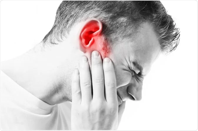
1. Ear infections:
What is Pneumatic Otoscopy?
In addition to standard otoscopy, there is a specialized technique called pneumatic otoscopy. This involves applying air pressure to the ear canal to evaluate the movement of the tympanic membrane.
Pneumatic otoscopy helps assess the functionality of the Eustachian tube, a small passage connecting the middle ear to the back of the throat. Problems with the Eustachian tube can cause issues such as fluid accumulation or middle ear effusion, hearing loss, or recurrent infections.2
Can You See the Eustachian Tube or a Cholesteatoma Via Otoscopy?
While otoscopy provides valuable information about the ear, it is important to note that it has its limitations. The Eustachian tube and cholesteatoma, an abnormal growth in the middle ear, are not directly visible through otoscopy alone.3 However, additional tests such as imaging studies or specialized procedures may be performed to further investigate and diagnose these conditions if they are suspected.
Summary
In conclusion, otoscopy is an essential diagnostic tool used to examine the ear and diagnose various conditions. Through otoscopic examination, healthcare professionals can identify ear infections, eardrum abnormalities, earwax impaction, and foreign objects. Pneumatic otoscopy aids in assessing Eustachian tube function.
Remember, otoscopy is a painless procedure, and it is typically performed by healthcare professionals such as otolaryngologists, primary care physicians, or audiologists.
References:
- National Institute on Deafness and Other Communication Disorders (NIDCD). (n.d.). Ear Infections in Children. Retrieved from https://www.nidcd.nih.gov/health/ear-infections-children
- Pneumatic otoscopy (2021). Mayo Clinic. Retrieved from https://www.mayoclinic.org/tests-procedures/pneumatic-otoscopy/about/pac-20385142 on 28 June 2023.
- Cholesteatoma (2022). MedlinePlus. Retrieved from https://medlineplus.gov/ency/article/001060.htm on 28 June 2023.
The information contained in this article is for educational and informational purposes only. You should not use the information as a substitute for, nor should it replace, professional medical advice. If you have any questions about your health, you should always consult with a physician or other health-care professional.
.png?branch=web_prod)

TR
Author
Tania Rodrigues, Audiologist
audiologist
Blog Article Carousel
The Blog Article Carousel allows you to showcase a selection of articles, and link to a Blog List or Aggregation page for more content.
 hearing and hearing loss
.png?branch=web_prod)