Overview:

The outer, middle and inner ear work together to create a hearing system that turns oscillations in the air into signals our brain can understand as sounds, speech and music.
The ear

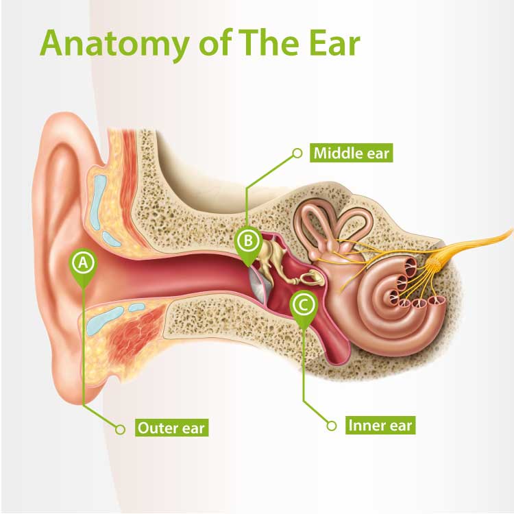
The structure of the human ear

The human hearing system consists of the outer ear, the middle ear and the inner ear.
 
The outer ear (auricle) that we commonly call our ear is only one part of hearing. The much bigger part of the ear is located in, and protected by, the skull.
 
The three parts of the ear are connected to each other via the ear canal. All acoustic systems run through it.
 
Tones, sounds and speech we hear are actually oscillations of the air. Before those oscillations are turned into sounds we can understand, they must pass from the outer to the inner ear via the middle ear - as well as all parts of our hearing system via the auditory nerve - to arrive in the brain as a signal.

How exactly does the ear work?

The outer ear
The Outer EarThe outer ear is formed by the area in front of the eardrum - the auricle and the outer ear canal. Sound waves arrive here and guided inwards through the auricle like a funnel.
 
The outer ear canal is shaped to: 
  • amplify the oscillating air that arrives, like in a resonator
  • improve our hearing by preventing wind and air movements from causing strong background noise.
The middle ear
The Middle EarThe middle ear is the area behind the thin membrane of the eardrum. When the eardrum is hit by sound waves, tiny oscillations reach the three little ossicles: the hammer, anvil and stirrup – the smallest bones in the human body.
 
Thanks to their unique location, these tiny bones can amplify the oscillations 20-fold, guaranteeing sound reaches the inner ear.

From the middle ear, the eustachian tube extends to the nose and throat area. This tube ventilates the middle ear and equalises pressure.
The inner ear
The Inner EarThe inner ear begins where the stirrup hits the next membrane – known as the oval window - which contains the organ of equilibrium and the cochlear.
 
The cochlear resembles a snail shell and is approximately the size of a pea. It contains three canals filled with a liquid.
 
The first canal directs the signals conducted into the liquid to the tip and back via a second canal. The central canal is home to the actual organ of hearing, the organ of Corti. The bottom is covered in thousands of tiny hairs.
 
The wave movements in the liquid-filled canals change depending on frequency. The hairs are only triggered by loud noises. The deeper the tones the further back in the cochlear the tiny hairs move, while high tones trigger the hair cells at the beginning of the cochlear.
 
Damage or “wear” of these tiny hairs in the cochlear is one of the main reasons for age related hearing loss.
How does the ear send a signal to the brain?
Even when sound waves arrive in the inner ear they still have no meaning. Once conducted to the auditory nerve, the sound waves are converted to electrical signals that first hit the brain stem.
 
The signal is conducted to the areas of the brain responsible for emotional assessment. So, meaning is attached to tones connected to existing patterns in the cortex. This enables us to understand speech, recognise the voice of a friend and judge dangerous situations.
 
This means our hearing only works if the signal is sent perfectly from one station to the other and only once oscillating air has been turned into a warning signal, pleasant music or a coherent sentence.

Air conduction versus bone conduction

Sound can reach the ear via air conduction or bone conduction.
 
In bone conduction, oscillating air hits the outside of the skull and makes it oscillate slightly. Conducted by the liquids in the ear, the oscillations reach the hair cells. Hearing via bone conduction is not as effective as air conduction hearing.

Did you know?

Bone sound wave conduction is the reason we find our own voice strange in recordings. You hear your own voice conducted via the air, instead of via the bones.